Epithelium
Lining epithelium
Simple squamous epithelium - endothelium
The slide contains dental pulp, pulpa dentis (HE). Under low magnification, one can see loose irregular connective tissue with numerous arterioles and blood capillaries, some of which filled with blood. Under high magnification, the nuclei of endothelial cells protruding into the lumen are visible in the walls of the blood capillaries.
The slide contains a kidney, ren (HE). Under low magnification, the cortex and medulla are visible. In the medulla, at higher magnification, collecting ducts (covered with simple cuboidal epithelium) are visible. In addition, we can see parts of the loop of Henle (covered with simple squamous epithelium) and blood capillaries (lined with endothelium).
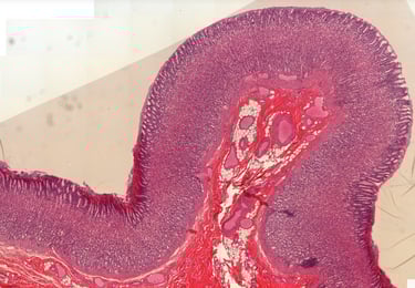
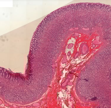
The slide contains a section of the small intestine, intestinum tenue (HE). The inner surface of the intestine is covered with a mucous membrane - mucosa, which consists of a lamina epithelialis made of simple cylindrical epithelium, a lamina propria (loose connective tissue) and a lamina mucularis mucosae (a circular layer of smooth muscle cells). At higher magnification, goblet cells - single-celled endo-epithelial glands - can be seen between the cylindrical cells of the epithelium (enterocytes). At high magnification, a striated border is visible on the apical surface of the cylindrical epithelial cells.
Pseudostratified cylindrical epithelium with cilia and goblet cells
The slide shows a longitudinal section through the respiratory part of the nose, respiratoria nasi (HE). The surface of the nasal conchae and nasal septum is covered with a pseudostratified columnar epithelium with the nuclei of columnar cells arranged in 2-3 rows. At higher magnification, cilia can be seen on the apical surface of the columnar cells, between which there are also goblet cells. At higher magnification, a thick basement membrane can be seen that separates the epithelium from the connective tissue on which it rests.
The specimen is the urinary bladder, vesica urinaria (HE). The inner lining of the bladder wall is covered with transitional epithelium. Cuboidal epithelial cells (with round nuclei) are arranged in several rows - this is a multi-row epithelium in which all cells are attached to the basal lamina by hemidesmosomes (these cannot be seen by light microscopy). Cells on the surface are narrow at the base, while their surface is convex, hence their name 'umbrella' cells. There is a crusta on their surface. The basal lamina between the epithelium and the connective tissue does not have connective tissue papillae.
Two-layered cuboidal epithelium
The specimen is a parotid gland, glandula parotis (HE). On the surface of the larger drainage ducts, in the connective tissue between the lobes, at high magnification, an epithelium made up of two layers of cuboidal cells can be seen.
Two-layered cylindrical epithelium
The specimen is a testis and epididymis, testis and epididymis (HE). The epididymal duct - ductus epididymidis - is lined with two layers of cells: cuboidal cells along the basal lamina and cylindrical cells on the surface. The superficial cells at the apical pole have large branched microvilli, called stereocilia.
Stratified squamous epithelium - non-keratinized
The preparation is the esophagus, oesophagus (HE). On the surface of the esophageal wall there is a multilayered, non-keratinized squamous epithelium. The epithelium is made up of a single layer of columnar cells along the basal lamina, a larger number of layers of cuboidal cells in the central part and a larger number of layers of squamous cells on the surface. The border between the epithelium and the connective tissue on which it lies (lamina propria) is irregular due to connective tissue papillae that push into the epithelium for increased contact strength.
Stratified squamous epithelium - keratinized
The specimen is skin, the integumentum commune (HE). The surface of the skin is covered by a multilayered, keratinized squamous epithelium. It is made up of a single layer of cylindrical cells along the basal lamina, a larger number of layers of cuboidal cells in the central part, and a larger number of layers of squamous cells on the surface. The squamous cells are covered by a thick layer of dead cells that exfoliate: the horny layer or stratum corneum. The boundary between the epithelium and the connective tissue on which it lies (lamina propria) is irregular due to connective tissue papillae that push into the epithelium.
Stratified cylindrical epithelium
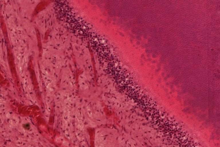
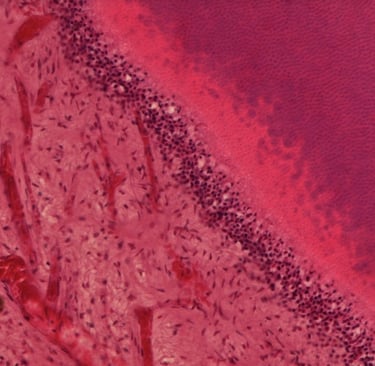
The slide shows a human embryo of approximately 8 weeks of age, sectioned across the abdomen. At higher magnification we see that the gastrointestinal tract, and particularly the stomach, has this rare form of stratified columnar epithelium.
Glandular epithelium
The specimen shows the pancreas, pancreas (HE). At low magnification, the lobes of the gland can be seen to be separated by connective tissue, which contain larger blood vessels and interlobular drainage ducts lined with double-layered cubic epithelium. The parenchyma of the gland is made up of serous terminal parts with a very narrow lumen, lined with pyramidal epithelial cells. The cells have a two-tone cytoplasm (darker at the base, lighter at the apex) due to the presence of zymogenic granules, and a large, round nucleus. In the center of the acinus are centroacinar cells. Between the terminal parts of the glands are intralobular drainage ducts lined with single-layered cubic epithelium. Within the parenchyma are the endocrine parts of the pancreas - the islets of Langerhans, built of strips of epithelial endocrine cells and blood capillaries.
The specimen shows female breast tissue, mamma (HE) in the active phase during lactation. The slide shows glandular lobules made of connective tissue with a large number of alveoli in which the secretion takes place. The glands are lined with a single-layer epithelium of varying height, which depends on the phase of secretion. The method of secretion of the breast is apocrine. The lobules are separated by dense connective tissue in which there are drainage channels - ductuli lactiferi lined with double-row cubic epithelium.
The specimen shows the skin, integumentum commune (HE). The surface of the skin is covered by multi-layered, keratinized squamous epithelium. The boundary between the epithelium and the connective tissue on which it lies (lamina propria) is irregular due to connective tissue papillae. There are several types of glands in the connective tissue under the epithelium. Sebaceous glands, gll. sebaceae are holocrine by secretion type. They are located next to hair follicles. Their terminal parts have a bag-like appearance, without a lumen that is filled with secrete and decayed cells. This type of gland is a polyptych - their terminal parts are lined with multiple layers of epithelial cells in different stages of decay.
Connective tissue
Blood and lymph
The specimen shows a blood smear (May-Grunwald-Giemsa). In addition to a large number of red blood cells (erythrocytes), white blood cells (leukocytes) can be seen: granulocytes (neutrophils, basophils, eosinophils) and agranulocytes (monocytes, lymphocytes). Individual clusters of blood platelets - thrombocytes can also be observed as small, dark eosinophilic spots. Erythrocytes are biconcave cells without a nucleus. Neutrophils are cells with a multi-segmented nucleus, eosinophilic cytoplasm, and hardly noticeable granules. Basophils are cells with a nucleus that is typically segmented in two sections, have an eosinophilic cytoplasm and blue-stained granules. Eosinophils are cells with a bi-segmented nucleus, eosinophilic cytoplasm, and red-stained granules. Monocytes are cells with a kidney-shaped nucleus and a basophilic cytoplasm that does not show granules. Lymphocytes are small cells that are hardly bigger than erythrocytes with a large round nucleus surrounded by a small rim of basophilic cytoplasm.
Connective tissue proper
Connective tissue proper is made up of three components: 1. a cellular fraction, 2. Fibers and 3. ground substance. The type and composition of cells can vary, but always includes fibroblasts. These cells are metabolically highly active, since they form the extracellular matrix. When these cells become dormant, they differentiate into fibrocytes which maintain the extracellular matrix. Fibers can be fibrous, elastic or reticular. Typically, the majority of fibers are formed by collagens. Ground substance is a gel-like tissue, made up of multi-adhesive proteins, glycoproteins, glycosaminoglycans, which through their structure and electrical charge, hold much water.
The specimen is a cross-section of the first 1/3 of the esophagus, oesophagus (HE). The slide shows that the wall of the esophagus is made up multiple layers. A thick layer of squamous, stratified epithelium can be seen on the surface of the tissue towards the lumen. Directly underneath, we find the lamina propria, which is made up of loose connective tissue. Since epithelium is avascular, the lamina propria is highly vascularized and feeds the epithelium. Loose connective tissue also contains a lot of ground substance, through which nutrients and gasses can easily diffuse to the epithelium. The lamina propria also provides a flexible structure and contains many immune cells, that can rapidlz neutralize micro-organisms that breach the epithelial barrier.
Dense irregular connective tissue
The specimen is skin with subcutaneous tissue, cutis and subcutis (HE) in the sole of the foot. Beneath the skin epithelium is a thin layer of loose connective tissue that enters the connective tissue papillae and is called the papillar dermis. Next, we see the thick, pink stained layer of the reicular dermis, which is made up of dense irregular connective tissue. It contains a dense network of mostly collagen type I fibers with very few cells.
Dense regular connective tissue
The specimen is a tendon, tendum (HE). On the longitudinal section, bundles of wavy collagen fibers are visible, which are all oriented in the same direction on order to be able to sustain maximum force rom one direction. Between the cells a very thin, loose connective tissue, called peritendineum internum, can be seen. Inside the bundles, between the individual fibers, we can see the elongated nuclei from tenocytes. Not visible on this specimen is the peritendineum externum, the outer layer of the tendon, consisting of loose connective tissue.
Connective tissue with special properties
The specimen is a mammary gland, mama (HE) in the inactive phase of an adult woman. The specimen shows lobules made of connective tissue with a large amount of white adipocytes separated by dense connective tissue. Adipocytes are characterized by a single (uniocular) vesicle filled with triglycerides. The cytoplasm and nucleus are pushed completely to the side. The tissue is sparsely vascularized and innervated and containes little connective tissue. The fat in the cells therefore gives the tissue its typical white-yellow color.
Supporting connective tissue
The specimen is a trachea (HE). This organ contains C-shaped plates of cartilage, localized under the submucosa. This is hyaline cartilage which has a layer of loose connective tissue or perichondrium surrounding the tissue. At higher magnification, isogenic groups of cells can be seen, consisting of chondrocytes located in lacunae surrounded by a layer of dark-colored intercellular matrix - the capsule. The space between the isogenic groups is a light-colored homogeneous matrix - the interterritory.
The preparation is an outer ear, auricula (HE). At the base of the ear there is an elongated plate of elastic cartilage surrounded by loose connective tissue of the perichondrium. At higher magnification, a network of purple/red-stained elastic fibers in the intercellular matrix can be observed between individual clusters of chondrocytes - isogenic groups.
The preparation is an ear, auricula (orcein). At the base of the ear there is an elongated plate of elastic cartilage surrounded by loose connective tissue of perichondrium. At higher magnification, a network of brown-colored elastic fibers in the intercellular matrix can be observed between individual clusters of chondrocytes - isogenic groups.
The specimen is part on an invertebrate disk (HE). This tissue consists of fibrocartilage, which is a mixture of hyaline cartilage and dense connective tissue. We can see chondrocytes as single cells, or grouped in small isogenous clusters in purple-stained cartilage. This tissue is inter spaced with pink-colored fibrous tissue in which we can find fibroblasts. These cells can be recognized by their more elongated nuclei. There is no distinct perichondrium. Fibrocartilage functions as shock absorber for bones such as the vertebrae
The specimen is a section of compact bone (ground, unstained). Osteons can be seen, which are formed by transversely cut Haversian canals surrounded by lamellae. In between lamellae are lacunae containing osteocytes. Individual Haversian canals are interconnected by transversely placed Volkmann canals. Only the inorganic part of the bone tissue is shown in the preparation. The canals and lacunae are empty because the cells were destroyed during preparation of the specimen.
The specimen is a cross-section of the diaphysis of a long bone (decalcification + HE). The diaphysis itself is made up of bone in which osteons can be observed: Haversian canals surrounded by lamellae with lacunae containing osteocytes. The lamellae are made up of thick collagen fibers that give them a homogeneous red color. The cavity of the diaphysis contains bone marrow. On the outer surface of the bone tissue is a layer of connective tissue - the periosteum. The inner lining of the bone is the endosteum. Under the periosteum and enosteum are the outer and inner general lamellae of bone tissue visible respectively.
The specimen is a longitudinally sectioned, decalcified long bone. This slide shows Haversian canals as elongated tubes, while Volkmann's canals are shown as interconnected tubes. Osteocytes are shown as elongated nuclei.
The specimen is a cross-section of the epiphyseal plate of a long bone (HE). The epiphyseal plate is typically lost in the second decade of life. The cartilage tissue, from which bone tissue develops, is divided into five zones (starting from the articular fissure): 1. Zone of reserve/resting - hyaline cartilage in which non-dividing chondrocytes are observed 2. Zone of proliferation - chondrocytes divide and specifically group together forming "columns" that are parallel to the longitudinal axis of the bone 3. Zone of hypertrophy - large, hypertrophic chondrocytes with a clearly increased amount of cytoplasm are visible. Resorption of the surrounding matrix is also observed. 4. Zone of calcified cartilage - characterized by two processes: gradual deterioration of chondrocytes and mineralization of the cartilage matrix. 5. Zone of ossification - islands of bone tissue surrounded by numerous blood capillaries and cells are observed.
The specimen is a longitudinal section of a long bone during embryonic development (HE). The cartilage tissue, from which bone tissue develops, is divided into five zones (starting from the articular fissure): 1. Zone of reserve/resting - hyaline cartilage in which non-dividing chondrocytes are observed 2. Zone of proliferation - chondrocytes divide and specifically group together forming "columns" that are parallel to the longitudinal axis of the bone 3. Zone of hypertrophy - large, hypertrophic chondrocytes with a clearly increased amount of cytoplasm are visible. Resorption of the surrounding matrix is also observed. 4. Zone of calcified cartilage - characterized by two processes: gradual deterioration of chondrocytes and mineralization of the cartilage matrix. 5. Zone of ossification - islands of bone tissue surrounded by numerous blood capillaries and cells are observed.
The specimen is stained with HE. In the deeper layers, ossification centers of different sizes can be observed. In them, osteocytes can be seen in lacunae, while at the edge of the ossification center there is one layer of cells made up of osteoblasts. In the loose connective tissue, which surrounds the already formed bone, there are numerous capillaries.
Muscle tissue
Smooth muscle
The specimen shows a cross-section of the stomach wall (HE). This organ has three layers of smooth muscle tissue, one longitudinal, one transverse and one oblique. At high magnification, the cells can be seen to be spindle-shaped in the longitudinal cut part with a centrally located nucleus. Cells are densely packed together and surrounded by a small amount of reticular connective tissue - the endomysium. In cross-sections, the cells have the shape of regular circles with or without nuclei, dependent on where the cell has been cut.
Skeletal muscle
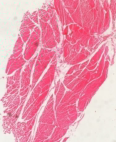
The specimen is a part of the tongue (HE). Skeletal muscle fibers are elongated cylinders in longitudinal section with a large number of nuclei located directly underneath the cell membrane. Around the cells is a reticular connective tissue - the endomysium, while around the bundles of muscle cells is a dense connective tissue - the perimysium. Cells in longitudinal sections show transverse striation. At high magnification, sarcomeres delimited by Z-lines, dark areas of A-stripes and light areas of I-stripes can be seen.
The specimen is a part of the tongue (HE). Skeletal muscle fibers are elongated cylinders in longitudinal section with a large number of nuclei located directly underneath the cell membrane. Around the cells is a reticular connective tissue - the endomysium, while around the bundles of muscle cells is a dense connective tissue - the perimysium. Cells in longitudinal sections show transverse striation. At high magnification, sarcomeres delimited by Z-lines, dark areas of A-stripes and light areas of I-stripes can be seen.
Cardiac muscle
The specimen is a part of musculature of the heart wall (HE), the myocardium. On a cross-section through the working musculature of the heart, irregularly shaped cardiac muscle cells with a centrally located nucleus surrounded by a large amount of soft connective tissue - endomysium can be observed. The bundles of muscle cells are surrounded by perimysium.
The specimen is a part of the musculature of the heart wall (HE), the myocardium. On a longitudinal section through the working musculature of the heart, cardiac muscle cells in the shape of elongated cylinders can be seen, which branch at the ends and connect with neighboring cells. The cells have centrally located nuclei (1-2) and are surrounded by a large amount of loose connective tissue - endomysium. At the point of connection of the cells, at high magnification, transitional plates or disci intercalares are visible as stepped darker lines. At higher magnification, the cells show transverse striation that is less visible than in skeletal muscle cells. Dark A-striations, light I-striations and sarcomeres can be seen.

Nervous tissue
Nervous tissue
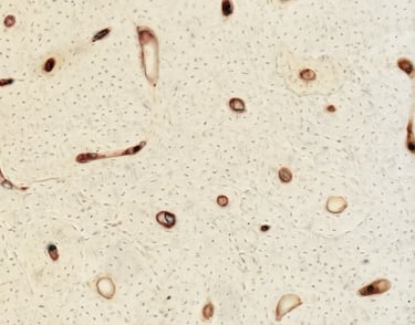
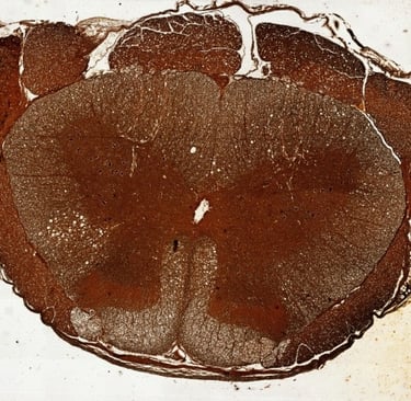
The specimen is the spinal cord, medulla spinalis (HE). On a cross-section of the spinal cord, the central canal can be seen. The central canal is lined by a single layer of cubic ependymal cells - specialized glial cells. Surrounding this canal an area of gray matter can be seen, while the white matter is located peripherally. In the gray matter, there are perikaryons of multipolar nerve cells, with a clearly visible nucleus and basophilic cytoplasm filled with Nissl bodies or tigroid (clusters of granular endoplasmic reticulum). Around the perikaryon is the neuropil, which consists of a network of nerve fibers, glial cells (microglia, astrocytes, oligodendrocytes) and blood capillaries. In the white matter area, we find myelinated nerve fibers, some glial cells and capillaries. Since myelin is dissolved during the preparation of the preparation, the fibers are surrounded by white space.
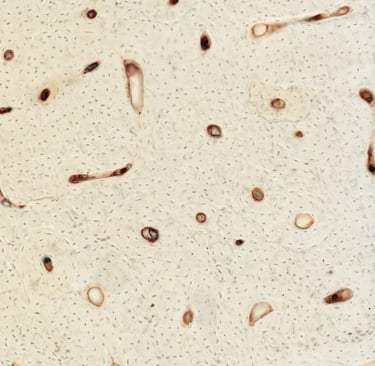
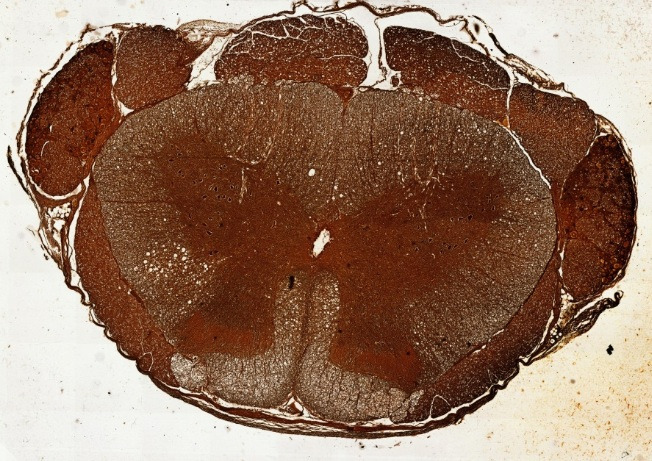
The specimen is the spinal cord, medulla spinalis (silver impregnation). On a cross-section of the spinal cord, the central canal of the spinal cord can be seen, surrounded by an area of gray matter, while the white matter is located peripherally. In the gray matter, there are darkly stained perikaryons of multipolar nerve cells with a light-colored nuclear area. Around the perikaryon is the neuropil, which consists of a network of black-stained nerve fibers, the nuclei of glial cells (microglia, astrocytes, oligodendrocytes) and blood capillaries. In the white matter area, we find myelinated nerve fibers, some glial cells and capillaries.
Follow
+385 51 651 176
Contact
Faculty of Medicine
Department of histology & Embryology
Braće Branchetta 20
51000, Rijeka, Croatia