General embryology
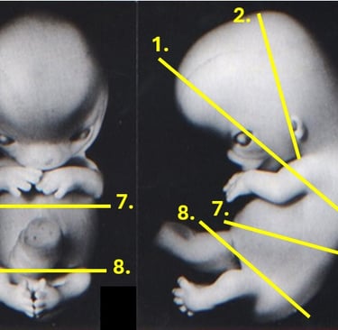
Sections through the human embryo (8-9 weeks)
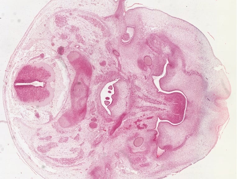
1. Section through the telencephalon and thorax
This section cuts through the front of the head and through the thorax. In the head, we see the two hemispheres (lateral ventricles) of the telencephalon. The section also goes through the eyes and the nasal cavity. The nasal cavity has started folding inwards, but has not yet formed the palatine shelf that will separate it from the oral cavity. In the center of the nasal cavity we see the cartilage of the septum. In the oral cavity, the tongue has formed ad we also see that the teeth have entered the early bell stage. In the lower part of the head we see the Meckel's cartilage around which the mandibles are being formed. In the upper thorax, we see the cartilage of the ribs and the upper limbs. We also see the upper pleura and two darkly stained parts of the descending thymus. In between the pleura, in a section of loose connective tissue, we see the aorta and primitive foregut. Towards the dorsal part, we see the cartilage of the vertebra surrounding a remnant of the notochord. Below this cartilage we see two sensory ganglia. Above the cartilage, we find two autonomous ganglia and the spinal cord, which is segregating in white matter on the outside and grey matter in the middle.
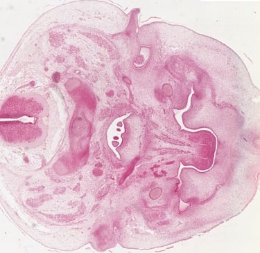
2. Section through the diencephalon and ear
This section cuts through the rear part of the head. We see the diencephalon of which the corpus striatum is bulging into the rapidly closing lumen. At the lower end we see the developing thalamus. In the lateral ventricles, we see the choroid plexus being formed. Below the developing brain, we see the inner, middle and outer ear being formed. On either side of the head, we see the otic vesicle. In this section, we see multiple vesicles, but these are connected. We also see the cartilage of the mallus and incus, which are formed from the first pharyngeal arch. The stapes is formed from the second pharyngeal arch and will join the other ossicles later. We also see the invagination of the external auditory meatus, which is formed from the 1st pharyngeal cleft.
This slide shows a section through the head of a human embryo. Moving from left to right, we first see the skin, which at this stage consists of a periderm covering a basal layer on top of the mesenchyme. Next we see the spinal cord, with very little white matter. We also see the autonomic ganglia, as well as the sensory ganglions. The tissue underneath, which will form the vertebrae, still contains a remnant of the notochord. IT has not yet differentiated into cartilage, which it will form before transforming into bone. Underneath, we see a small section of the nasal cavity and the tongue with salivary glands. On either side we also see the circular shape of Meckel's cartilage, which guides the formation of the mandibulae.
This slide shows an embryo one to two weeks older than the previous one. We can see that the spinal cord has formed more white matter. We can also see that the vertebrae are now made entirely of cartilage, although the notochord can still be seen. Below, the esophagus and aorta can be found lying side by side. We also see the large lumen of the primary bronchi and lungs on either side, which do not contain alveoli. We also see the heart, which is relatively large at this stage. We can clearly see both the atria and ventricles. The pleural cavity and pericardium have already separated. Behind the pleural cavity, we see the formation of ribs, which are still cartilaginous. Finally, we see one of the upper limbs.
5. Section through the upper abdomen
In this preparation, the embryonic liver dominates, which is relatively large at this stage. It should be noted that this organ contains many small cells, which are cells from the hematopoietic lineage. At the bottom of the preparation we see the umbilical cord.
6. Section through the lower abdomen (8 weeks)
In this slide we can still see the lower part of the liver. In addition, beneath the cartilage of the forming vertebrae we see the metanephros on either side of the specimen, which are ascending through the embryo. Note the S-shapes tubules, which will differentiate into the proximal and distal tubules and the loop of Henle. Below that we find two dense structures, which are the gonads, still conneccted to a remnant of the mesonephros. We also see two tubes with convoluted epithelium that are the developing small intestine. Between these two tubes we see the developing pancreas. The tube with cilindrical, nonconvoluted epithelium is the stomach. Below the embryo we see the umbilical cord.
7. Section through the lower abdomen (9 weeks)
This specimen is about two weeks older than section 6. We now see that the metanephros has formed glomeruli and we see differences in the epithelium of the proximal and distal convoluted tubules and we see a clear medulla. The pancreas has further expanded, even though it is still in the pseudoglandular stage. The epithelium of the small intestine has further convoluted. At the ventral side of the embryo, we see the umbilical vein carrying oxygenated blood from the placenta entering the embryo.
8. Section through the pelvic region
This section of the pelvic region shows the urogenital ridge, which has mostly separated in a gonadal ridge and mesonephros. The large, canaliculized structures are the two mesonephri. These structures will disappear by the 10th week of development. The dense lobes attached to these structures are the developing gonads. Finally, we see parts of the developing hindgut. This specimen is 7-8 weeks of age and the vertebrae have not formed cartilage yet.
Embryonic tissue
At the bottom of the slide, near the spinal cord, we see the mesenchyme. This tissue resembles loose connective tissue, as it contains much ground substance and few cells. It is a loosely organized, mostly mesodermal-derived embryonic tissue that develops into connective and skeletal tissues, including blood and lymph.
The specimen is the umbilical cord, chorda umbilicalis (HE). The umbilical cord contains two arteries with thick walls and regular lumen and one vein with irregular lumen. Between these structures we see Wharton's jelly. This tissue resembles loose connective tissue, as it contains a lot of ground substance and few cells. Its function is to absorb pressure on the umbilical cord to ensure that the blood vessels remain open even when the baby compresses this organ.

Extra-embryonic structures
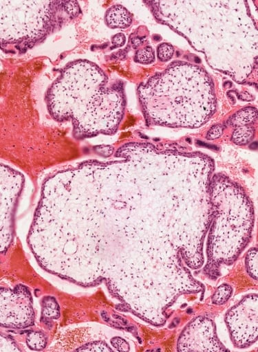
Chorionic vili (tertiary)
The specimen is a part of the placenta with cross-sectioned chorionic villi, chorion villi (HE) surrounded by maternal blood. The interior of the villi is made up of mesenchyme with blood capillaries containing fetal blood. The surface of the villi is initially covered by two layers: an outer syncytiotrophoblast and an inner cytotrophoblast. At later stages, the cytotrophoblast is mostly lost, which has occured in most vili of this specimen. Placental fibrinoid, an eosinophilic mass that thickens the placental barrier, is deposited on the surface of the chorionic villi.
The specimen is the umbilical cord, chorda umbilicalis (HE). The umbilical cord contains two arteries with thick walls and a circular lumen and one vein with a large, irregular lumen. The section shows the allantois, an irregular tubule covered with cubical epithelium, which will be lost at later stages of development. Between these structures we see Wharton's jelly, which surrounds the blood vessels and the allantois, providing them with mechanical protection. The surface is covered by epithelium of the amnion.
Development of the eye
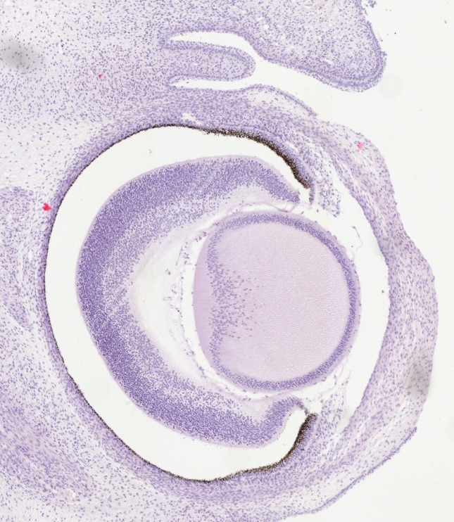
Development of the eye - I early phase
The specimen is a section through part of the fetal head (HE). Immediately below the surface epithelium, the basis for the development of the retina of the eye is visible - the optic cup. The optic cup has two layers - the outer layer is made up of a single layer of pigmented cells - stratum pigmenti retinae (in the larger posterior part), stratum pigmenti corporis ciliaris (the smaller, anterior, serrated part) and stratum pigmenti iridis (the anterior part that passes into the inner layer in the central part of the eye pitcher). In this specimen it can be seen as a layer of cilindrical cells with brown granules in their cytoplasm, especially towards the apical side. The inner layer is a cluster of cells that provides the basis for the retina: in the anterior part it is thinner - the basis for the pars iridica retinae - the anterior pigmented layer to which the unpigmented pars ciliaris retinae continues. Next, we see a thick layer of cells from which the pars optica retinae will develop. The place where the optic nerve exits is visible at the back of the optic cup of the eye on the right side of the specimen. The lens - lens cristallina is visible in the concavity of the optic cup on the left side. The mesenchyme around the optic cup begins to form the middle and outer eye layers that will later differentiate into the fibrous and vascular layers. The basis from which the eyelids will gradually develop is also visible.
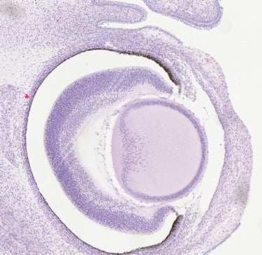
Development of the eye II - intermediate phase
The slide is a section through part of the fetal head (HE). At this stage we see that the lens has further developed, with fibers covering almost the entire center of the organ. We also see the further formation of the eyelids and conjunctiva.
Development of the eye III - late phase
The section is a cross-section through part of the fetal head (HE). In this specimen, the anterior chamber of the eye is visible between the base of the lens and the base of the cornea. Since the eyelids have fused at this stage of development, the space lined by the conjunctiva can be seen between the outer surface of the cornea and the inner surface of the eyelids. The outer vascular layer can now also be seen, as well as the onset of the outer fibrous layer.
Development of the teeth
Development of the teeth I - early phase
The specimen is a section through part of the fetal head (HE). 7-8 weeks after fertilization, teeth appear as dental buds that branch from the dental lamina in the oral epithelium. In this slide, we see how this bud initiates condensation and differentiation of the underlying tissue, which will form the dental papilla. We also see how the vestibular lamina branches off as a separate bud from the oral epithelium
Development of the teeth II - early phase
The specimen is a section through part of the fetal head (HE). This section was taken just a few days later than the previous slide. The orientation of the cut is different, which is why we see the developing brain in this slide. We can see that the nasal cavity is expanding and the maxillary bone is forming by intramembranous ossification. The tooth bud is expanding further into the underlying ectomesenchyme.
Development of the teeth III - Early cap phase
The specimen is a section through part of the fetal head (HE). The slide shows how the oral epithelium starts folding around the underlying tissue, which will differentiate into the dental papilla. At the upper jaw, we see that the dental bud is still expanding into the underlying tissue and did not form a cap yet. We do see further segregation from the vestibular lamina. In the lower jaw, we see that the cap is being formed.
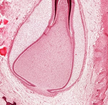
Development of the teeth IV - Late cap phase
The specimen is a section through part of the fetal head (HE). We are now several weeks later in the third month of development. We see that the dental epithelium has separated from the oral epithelium and lies as a cap on the dental papilla, a structure known as the enamel organ. The cap covers the dental papilla and is filled with stellate reticulum. At the interphase of these two tissues the underlying mesoderm will differentiate into odontoblasts. The lower end of the enamel organ, formed by the inner enamel epithelium, will differentiate into ameloblasts. We can also see that the cervical loops are expanding downward, which will form the epithelial root sheath.
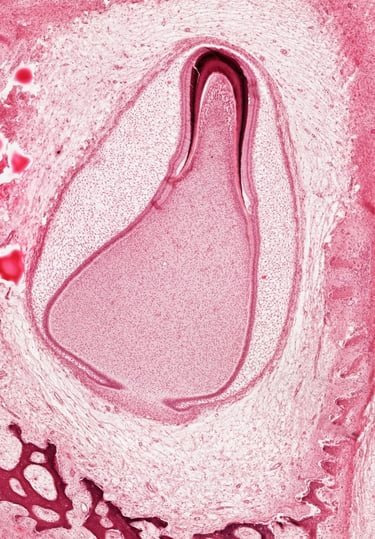
Development of the teeth V - Early bell phase
The specimen is a section through part of the fetal head (HE). At approximately six months after fertilization, we see that the shape of the lower jaw is clearly visible, with hairs developing in the outer skin. Around the teeth, the stellate reticulum covered by the dental epithelium now completely surrounds the dental papilla and has become very thin at the place where tissue has calcified. The odontoblasts have formed dentin, which can be seen as a dark red/pink layer overlying a lighter pink stained layer pf predentin. Surrounding this layer, we see a black layer of enamel, formed by ameloblasts, the cylindrical cells that can be seen lining this tissue. Over the ameloblasts is the stratum intermedium, which helps with calcification of tissue. At the lowr part of the tooth, the Hertwig's epithelial root sheath (HERS) has formed and folded inwards, defining where later the apical foramen will be.
Follow
+385 51 651 176
Contact
Faculty of Medicine
Department of histology & Embryology
Braće Branchetta 20
51000, Rijeka, Croatia